Regional differences
Which expression patterns distinguish different tissue regions?
ZYENRA-SPATIAL V2 · SPATIALLY BARCODED CHIP
Connect molecular signals, coordinates and tissue images with spatial barcodes to investigate expression patterns across tissue regions.
Current scope: fresh-frozen, OCT-embedded tissue. Pre-fixed tissue and FFPE are not currently accepted; spatial RNA services are under optimization and assessed per project.
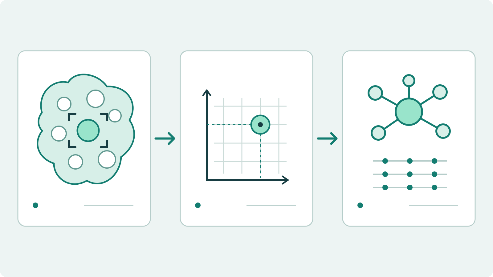
How tissue location, spatial coordinates and molecular information connect.
RESEARCH PERSPECTIVE
Which expression patterns distinguish different tissue regions?
How do molecular signals relate to morphological boundaries and tissue structure?
How can tissue position inform research into molecular signals within local cellular environments?
HOW IT WORKS
01
Barcode information on the chip provides the basis for spatial association.
02
Retain tissue morphology through imaging for subsequent spatial alignment.
03
After sequencing, link expression information to spatial barcodes and analyze it in the context of tissue images.
V2 CHIP FORMATS
Two layouts share a 25 × 75 mm slide format. The diagrams illustrate the layouts; experimental configuration is confirmed for each project.
FORMAT A
Two areas, each measuring 15 × 5.5 mm.
165 mm²
Total capture area
FORMAT B
One area measuring 10 × 10 mm.
100 mm²
Total capture area
ZYENRA-spatial v2 format parameters; effective resolution depends on tissue, diffusion and analysis settings.
WORKFLOW
Start with fresh-frozen, OCT-embedded tissue and assess morphology, RNA quality and sectioning plans.
Perform section fixation, tissue imaging, permeabilization and capture according to the workflow.
Use the assay reactions and external sequencing to obtain molecular and barcode information.
Combine spatial barcodes and tissue images to study regional expression and the relevant quality metrics.
SAMPLES & SCOPE
Fresh-frozen, OCT-embedded tissue. Discuss a pilot study around tissue morphology, RNA quality, dimensions and sectioning conditions before submission.
Sample requirements and in-workflow fixation are separate: submitted tissue must be unfixed and fresh-frozen, while the workflow itself fixes sections after mounting. Section fixation within the workflow does not imply compatibility with pre-fixed input. The current workflow does not accept pre-fixed or paraffin-embedded samples.
Confirm regions of interest and sample compatibility before planning sectioning, imaging, experiments and analysis. Spatial RNA services are under optimization and assessed project by project.
Feedback, suggestions or concerns: CEO Direct